|
 |

 A neurologically intact patient with a deep tumor
that is poorly defined by MRI or CT is the ideal candidate for
stereotactic biopsy. Such procedures are also
useful for patients with recurrent tumors in whom a change in
histopathology is anticipated and when the use of interstitial
irradiation or hyperthermia is planned. Some
patients with cystic recurrences obtain symptomatic relief from
stereotactic aspiration of the cyst. The advent of computerized
imaging and CT - and MRI -compatible stereotactic frames (e.g.,
the Leksell instrument) has greatly simplified the performance
of stereotactic procedures for both large and small target
lesions. Nevertheless, simple biopsy carries less than a 2
percent mortality rate and a 3 percent serious complication
rate. Although the smear preparations from such procedures yield
a correct diagnosis in less than 95 percent of glial tumors when adequate
tissue has been obtained, in 11.8 percent of the cases either
the diagnosis is incorrect or the material is inadequate.
After stereotactic biopsy, all patients with a confirmed
diagnosis of a glial neoplasm should undergo some form of
external irradiation. Combined stereotactic biopsy and
postoperative irradiation is an especially appropriate method
of handling young and neurologically intact patients with
large, non enhancing, low-density lesions on CT or ill-defined, nonenhancing lesions on MRI. Even patients with high-grade
tumors can do well with such management. In cases of diffuse
spread of a low-grade astrocytoma through large volumes of
critical tissue, it is usually impossible at open craniotomy to
do more than a biopsy, because the margins of the lesion are
totally undefined. Because open biopsy without resection carries
a higher complication and mortality rate than either closed
biopsy or radical removal, it seems only prudent to subject such
patients to stereotactic biopsy instead.
A neurologically intact patient with a deep tumor
that is poorly defined by MRI or CT is the ideal candidate for
stereotactic biopsy. Such procedures are also
useful for patients with recurrent tumors in whom a change in
histopathology is anticipated and when the use of interstitial
irradiation or hyperthermia is planned. Some
patients with cystic recurrences obtain symptomatic relief from
stereotactic aspiration of the cyst. The advent of computerized
imaging and CT - and MRI -compatible stereotactic frames (e.g.,
the Leksell instrument) has greatly simplified the performance
of stereotactic procedures for both large and small target
lesions. Nevertheless, simple biopsy carries less than a 2
percent mortality rate and a 3 percent serious complication
rate. Although the smear preparations from such procedures yield
a correct diagnosis in less than 95 percent of glial tumors when adequate
tissue has been obtained, in 11.8 percent of the cases either
the diagnosis is incorrect or the material is inadequate.
After stereotactic biopsy, all patients with a confirmed
diagnosis of a glial neoplasm should undergo some form of
external irradiation. Combined stereotactic biopsy and
postoperative irradiation is an especially appropriate method
of handling young and neurologically intact patients with
large, non enhancing, low-density lesions on CT or ill-defined, nonenhancing lesions on MRI. Even patients with high-grade
tumors can do well with such management. In cases of diffuse
spread of a low-grade astrocytoma through large volumes of
critical tissue, it is usually impossible at open craniotomy to
do more than a biopsy, because the margins of the lesion are
totally undefined. Because open biopsy without resection carries
a higher complication and mortality rate than either closed
biopsy or radical removal, it seems only prudent to subject such
patients to stereotactic biopsy instead.
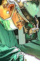
A recent development has been the evolution of
hybrid or combined techniques in which the precision and
accuracy of imagebased stereotaxy is combined with the
therapeutic advantages of open procedures. Patients are placed
into a stereotactic frame and targets are calculated from an
enhanced MRI or CT scan in the usual fashion. Upon return to
the operating room, a stereotactic probe is used to guide the
placement of the scalp incision and bone flap. After these have
been turned, the probe is again lowered to the surface of the
operative field and the dural incision selected. Finally, the
probe can be used to guide the placement of the cortical
incision and in some instances can be followed all the way down
the transcortical tunnel until the tumor is reached. This
method obviously avoids virtually all possible errors of localization
in the management of deeply placed and ill-defined
small lesions, Frameless stereotaxy is also in use as
an intraoperative aid for craniotomy. In this method, a
robotic arm is touched to the patient's head and acquires
localization points for a computer in which the image of the
lesion has been previously stored, Stacked slice representations
of the tumor within the brain have also been used to perform
computer-guided stereotactic resections of deep lesions by
mounting a laser and computercontrolled motors on a
stereotactic frame.
 
|
|
|
|
|
This site is non-profit directed
to medical and neurosurgical audience to share
problems and solutions for brain tumors
diagnosis and treatment modalities.
Author of the
site.

Prof. Munir A. Elias MD., PhD.
Facts of life
When entering the soul of the human, there is a
great discrepancy about the value of timing of
the life. Some are careless even about the
entire of their existence and others are
struggling for their seconds of life.
Quality of life
It plays a major impact in decision making from
the patient. Here come the moral, ethics,
religious believes and the internal motives of
the patient to play a major hidden role in his
own survival.
|
|
|