Colloid Cysts
occur infrequently and
account for less than 1 percent of intracranial tumors.
However, there is a suggestion of an increased rate of
detection coincident with improved neuroimaging techniques.
They can produce symptomatic obstruction of the foramina of
Monro and have been implicated as a cause of sudden
deterioration and death, although their natural history is
not well understood. They also may occur without causing
symptoms and may appear as incidental findings at autopsy.
Colloid cysts are round or
oval and vary in size from a few millimetres to several
centimetres in diameter. The site of origin is most commonly the
roof of the third ventricle at the level of the foramina of
Monro. Such cysts are usually homogeneous and hyperdense on non
contrast CT and show minimal enhancement after contrast
administration. MRI shows similar characteristics (Fig.1). Small
colloid cysts may be found incidentally; if this is the case and
they are not causing problems, the patient should be followed
carefully because the cysts can and do grow. Patients with
symptomatic colloid cysts have two common clinical
presentations. In the younger group, less than 40 years of age,
the usual problem is unlocalized increased intracranial pressure
in association with moderate hydrocephalus. A story of
intermittent obstruction of the foramina of Monro causing
headaches which are relieved by changes in head position seems
to be overemphasized.
Likewise the incidence of
sudden death due to an abrupt and complete blockage of CSF flow
by these cysts is overstated. The cysts are not adherent to the
foramen of Monro, are not very mobile, and rarely completely
occlude the foramen. In most cases of sudden death there is a
history suggestive of increasing intracranial pressure for weeks
to months. Complete occlusion is obviously not necessary to
interfere with CSF flow enough to cause hydrocephalus. In older
patients, dementia and hydrocephalus without increased
intracranial pressure can be seen.
The optimal surgical
management of colloid cysts remains controversial. Surgical
strategies, include a transcortical transventricular approach
through a craniotomy, a transcallosal approach via a craniotomy,
and a stereotactic craniotomy approach. Needle aspiration of the
cyst contents has been proposed as an alternative to craniotomy.
The various techniques for evacuating cyst contents include
freehand aspiration, stereotactic aspiration, CT -assisted
stereotactic aspiration and endoscopic aspiration. Shunting of
one or both lateral ventricles may be useful in certain
instances, but is considered to be the least satisfactory of all
methods for dealing with colloid cysts.
Problems associated with cyst
aspiration include difficulty penetrating a thick cyst wall,
difficulty aspirating viscous cyst contents, and the possibility
of neural or vascular injury if the procedure is not visualized
by the surgeon. Failure of stereotactic cyst aspiration has been
correlated with a hyperdense appearance on the preoperative CT
scan. Recently, a high recurrence rate has been reported
following aspiration of colloid cysts.
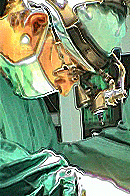
A rigid endoscope and its
sheath are introduced into the lateral ventricle through a
coronal burr hole, The colloid cyst is identified at the foramen
of Monro and the cyst capsule is opened using a fiberoptic
Nd:YAG laser introduced through a working channel in the
endoscope sheath. Frequently small vessels are seen draped over
the tumor capsule and these can be cauterized with the laser
prior to opening the cyst. The cyst contents are then emptied
with a suction catheter introduced through a working channel in
the endoscope sheath. If the cyst contains solid material, this
can be removed using grasping forceps, Once the cyst is emptied,
the remaining capsule is coagulated and shrunk with the laser
and then removed in a piecemeal fashion using microscissors and
grasping forceps, The septum pellucidum can be fenestrated using
the laser. The ventricular system is irrigated copiously and a
ventricular drain is left in place for 48 h in an attempt to
clear the irritative cyst contents. In addition, dexamethasone
is administered perioperatively.
The ventriculoscope is useful
in treating colloid cysts because it allows the operator several
options. If after inspection the surgeon thinks that the cyst
cannot be removed endoscopically, then either the cyst may be
removed via a craniotomy or the septum may be fenestrated
endoscopically and a ventricular shunt inserted, In medically
unstable patients, endoscopic cyst evacuation may be performed
under local anaesthesia.
Both groups of patients
respond well to treatment of the hydrocephalus which is best
accomplished by removing the colloid cyst. This can be done in a
variety of ways, but it seems that the endoscopic approach may
evolve as the least invasive and most beneficial way. In cases
in which cyst removal is not a reasonable option for whatever
reason, the hydrocephalus can be treated with a shunt. Each
lateral ventricle may require shunting individually; however,
this has not commonly been required because there is usually a
defect in the septum pellucidum.